Metabolic Dysfunction-Associated Steatosis Liver Disease (MASLD)
Metabolic Dysfunction-Associated Steatosis Liver Disease (MASLD, previously referred to as NAFLD, non-alcoholic fatty liver disease) is caused by an abnormal amount of fat buildup in the liver not due to alcohol consumption.
MASLD versus MASH:
- MASLD: fat buildup in the liver, but little or no swelling and cell damage. MASLD can be diagnosed with imaging such as an ultrasound, transient elastography (Fibroscan), MRI, or CT.
- MASH, Metabolic Associated Steatohepatitis: fat buildup in the liver with swelling and cell damage. MASH can only be diagnosed with a liver biopsy.

The longer the amount of time that high levels of steatosis or fat are present on your liver, the higher the chances it will be converted into scarring. Therefore, if left untreated, MASLD can lead to complications such as scarring on the liver (fibrosis), cirrhosis, liver cancer, and other health problems. It is still unclear in the medical field as to why some individuals have MASLD, while others will develop into MASH.
MASLD affects one in three Americans. Both MASLD and MASH can be reversed, which elevates the need for clinical care and supports the promise of research advancements.
Common Symptoms of Advanced Liver Disease
- Fatigue
- Nausea
- Unexplained weight loss
- Loss of appetite
- Abdominal pain, especially on the right side
- Swelling in the legs
Preventative Measures
- Aim to have a healthy diet consisting of more unprocessed foods than processed. Adopt diets like the Mediterranean diet which is rich in nutrients.
- Maintain a healthy body weight.
- Try to exercise often, about 30-60 minutes 2-3 times a week.
- Cut back on smoking and drinking.
- Lessen the amount of sugary and alcoholic beverages consumed.
Contributing Factors
- Type 2 diabetes
- Obesity, especially with a large waist size
- Insulin resistance or prediabetes
- High cholesterol (mainly LDL or “bad” cholesterol) or triglyceride levels
- High blood pressure
- Sleep apnea
- Polycystic ovarian disease
Learn more about the imaging that helps diagnose MASLD
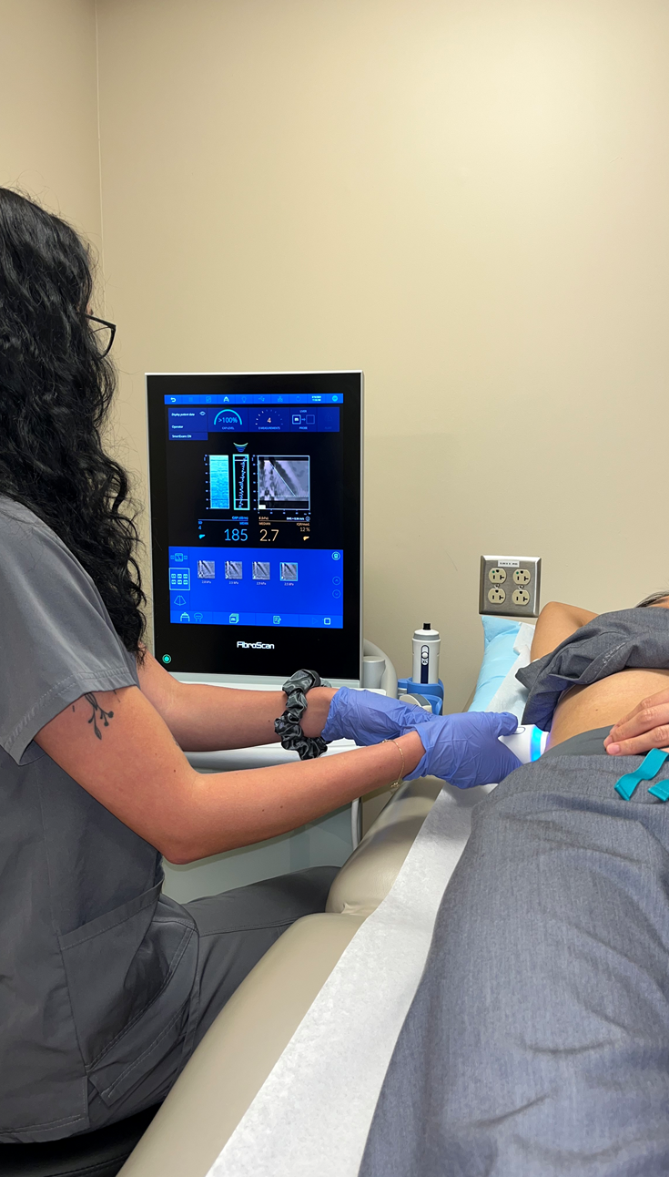
Transient Elastography
Transient Elastography (also referred to as a fibroscan) is a noninvasive imaging technique used to measure the amount of steatosis or fat present on your liver and how stiff it is (or how much fat has been converted to scarring).
The generated CAP number (the whole number) will indicate steatosis present on the liver. The higher the CAP number, the higher the presence of steatosis.
The kPa number (the number displayed as a decimal point) indicates the stiffness or scarring on the liver. The higher the kPa number, the higher the presence of scarring.

Ultrasound Elastography
Shear-Wave Ultrasound Elastography (SWE) is a noninvasive imaging technique that uses shear waves to determine the stiffness of internal organs. The exam will produce an LSM mean (liver stiffness measurement mean). The faster speed of shear waves indicates less stiffness of an organ, or less fibrosis (scaring).

Magnetic Resonance Elastography
Magnetic Resonance Elastography (MRE) is a noninvasive imaging technique used to help determine the stage of liver disease. It is a preferred method as it shows the entire structure of the liver and its stiffness. The exam is done by placing an MRE pad over a patient’s gown on the abdomen where it sends vibration waves through the liver during an MRI.
The exam produces a color-coded picture like the MRE provided, indicating ranges with blue as healthy soft tissue and red as stiff tissue or fibrosis. The exam will also generate a Mean LSM: <2.5 kPa = Normal, 3.0 to 3.5 kPa = Stage 1-2 fibrosis, 3.5 to 4.0 kPa = Stage 2-3 fibrosis, 4.0 to 5.0 kPa = Stage 3-4 fibrosis, 5.0 kPa = Stage 4 fibrosis or cirrhosis.
Computed Tomography
Computed Tomography (CT) is a noninvasive imaging technique that uses X-rays and computer technology to produce images of the body at multiple angles. Unlike an X-ray, CT scans can show detailed images of bones, muscles, fat, and organs.

Liver Biopsy
A liver biopsy is an invasive technique performed to diagnose many liver diseases and lesions as well as to assist in determining a fibrosis score.
A tiny portion of liver tissue is removed, via a hollow needle, which will be used to represent the condition of the entire liver. Before this test, an ultrasound or CT may be performed to determine the needle insertion spot.
There are multiple ways to perform a liver biopsy. One way is the percutaneous biopsy, where a needle is inserted through the abdominal skin and directly into the liver. Percutaneous is the preferred choice, as it is the least invasive biopsy method. Another way is the transjugular biopsy, where a needle is inserted into a catheter and travels down the jugular vein to obtain a sample from the liver. This method may be preferable for a patient if there are concerns surrounding bleeding and clotting during a percutaneous biopsy.


Histology of a Liver Biopsy
A pathologist studies the liver biopsy sample to look for indications of disease or damage. Certain patterns on the sample will indicate inflammation, steatosis, fibrosis, or a tumor. For example, the picture to the left shows a blue chicken wire pattern representing fibrosis.
The pathologist will then send a report of what they observed on the sample to the healthcare provider, stating possible diseases or fibrosis stage.
